Marburg Virus Under Microscope - Antiviral Aims To Prevent Spread Of Deadly Marburg Virus Technology Networks / The world health organization (who) rates it as a risk group 4 pathogen.
Marburg Virus Under Microscope - Antiviral Aims To Prevent Spread Of Deadly Marburg Virus Technology Networks / The world health organization (who) rates it as a risk group 4 pathogen.. There are four known strains of ebola virus but only one strain of marburg virus. Diese pandemie kam nicht zufällig zustande. In addition, exposure to an infected human is high risk factor. When seen under a microscope. Click link below to learn how to optimize your immune.
The intelligent design of the immune system. People infected with the virus suffer from tissue inflammation, sepsis and skin bleeding. Filovirus means thread virus and refers to the way the virus looks under a microscope. He says the vaccine approach can be extended to other members of the same virus family, such as marburg virus, which is also a major threat. The marburg virus contains seven structural proteins.
Marburg Virus Images Stock Photos Vectors Shutterstock from image.shutterstock.com Marburg virus is the causative agent of marburg virus disease (mvd), a disease with a case fatality ratio of up to 88%. The marburg virus under a microscope. The intelligent design of the immune system. If you look at the history of marburg and ebola outbreaks, there are several links between these diseases and bats. When seen under a microscope. The junin virus is associated with argentine hemorrhagic fever. People infected with the virus suffer from tissue inflammation, sepsis and skin bleeding. Get a microscopic look of what happens daily in your body.
It is unknown how marburg virus first transmits from its animal host to humans;
Electron microscopic studies following the 1975 marburg virus infections in south africa were limited to cell culture preparations and liver specimens from the single fatal case (cdc and south african institute for medical research, johannesburg). The marburg virus is part of the filovirus family (1). The junin virus is associated with argentine hemorrhagic fever. Murphy shot some images of the virus in the microscope. Marburg virus is the causative agent of marburg virus disease (mvd), a disease with a case fatality ratio of up to 88%. Marburg virus is a deadly pathogen that causes marburg disease a severe viral hemorrhagic fever, named after the city in germany, where the first outbreak occurred in 1967. The problem is that the symptoms can appear to be so common that the disease is rarely detected or identified in the first instance. Risk factors include exposure to african green monkeys and certain bats; The intelligent design of the immune system. When seen under a microscope. These types of viruses encode their genome in the form of single stranded negative polarity rna. Therefore, we can not see viruses under the microscope. Once the virus infects a human, the infections can spread from person to person.
Marburg virus under the microscope. Murphy shot some images of the virus in the microscope. People infected with the virus suffer from tissue inflammation, sepsis and skin bleeding. After incubating for five to 21 days, the disease comes on suddenly with symptoms including fever, chills, headache and. Get a microscopic look of what happens daily in your body.
A Marburg Virus Treatment Approach Is Identified Microbiology from d3bkbkx82g74b8.cloudfront.net The size of viruses ranges from 20 to 400 nm, which is too small to be seen with an optical microscope. When seen under a microscope. After incubating for five to 21 days, the disease comes on suddenly with symptoms including fever, chills, headache and. Marburg virus under the microscope. Murphy shot some images of the virus in the microscope. The marburg virus under a microscope 6. Marburg and ebola viruses are both members of the filoviridae family (filovirus). It is unknown how marburg virus first transmits from its animal host to humans;
Viruses under the microscope virions are very small ranging between 400nm (mimivirus, poxviruses) to about 25nm (polio virus) given that the resolution of conventional compound microscopes is limited to half the wavelength of radiation that is typically used for imaging (200 nm) they only allow for users to view the general morphology of giant viruses.
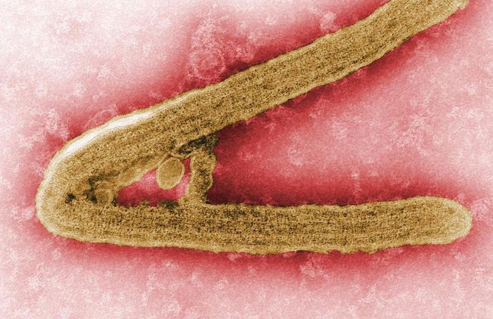
At the time, similarly afflicted patients were identified in belgrade. By fiona zerbst in conversation with prof robert swanepoel. Murphy in 1968, depicts a marburg virus virion, which had been grown in an environment of tissue culture cells. Filovirus means thread virus and refers to the way the virus looks under a microscope. In addition, exposure to an infected human is high risk factor. People infected with the virus suffer from tissue inflammation, sepsis and skin bleeding. Those pictures are still the most commonly available images of the virus, including the photo included in this post and a brightly colored. The genus marburgvirus derives its name from the city in germany that was one of the sites where infection with the virus was initially identified (reviewed in ref. Ebola and marburg viruses (marv) are members of the filoviridae and induce a serious hemorrhagic disease. Marburg virus is the causative agent of marburg virus disease (mvd), a disease with a case fatality ratio of up to 88%. Marburg virus under the microscope. Marburg virus disease is caused by viruses that produce symptoms of fever, chills, headaches and muscle aches early in the disease; These types of viruses encode their genome in the form of single stranded negative polarity rna.
Marburg virus is a deadly pathogen that causes marburg disease a severe viral hemorrhagic fever, named after the city in germany, where the first outbreak occurred in 1967. Diese pandemie kam nicht zufällig zustande. Those pictures are still the most commonly available images of the virus, including the photo included in this post and a brightly colored. Monath in the light of progress in virology, it comes as a surprise to discover a disease, an epidemic disease moreover, caused by a new virus. Get a microscopic look of what happens daily in your body.
Persistent Marburg Virus Infection In The Testes Of Nonhuman Primate Survivors Sciencedirect from ars.els-cdn.com The virus is considered to be extremely dangerous. The intelligent design of the immune system. If you look at the history of marburg and ebola outbreaks, there are several links between these diseases and bats. The authors began by isolating marburg virions from infected cells, and imaging them using cryoem, a technique that freezes samples to prevent their distortion in the evacuated atmosphere of the electron microscope. When seen under a microscope. Marburg virus is a deadly pathogen that causes marburg disease a severe viral hemorrhagic fever, named after the city in germany, where the first outbreak occurred in 1967. Marburg virus disease is caused by viruses that produce symptoms of fever, chills, headaches and muscle aches early in the disease; The world health organization (who) rates it as a risk group 4 pathogen.
Ben couwenberg now i posit he was under hyperspacial microscope.
At the time, similarly afflicted patients were identified in belgrade. Murphy in 1968, depicts a marburg virus virion, which had been grown in an environment of tissue culture cells. Marburg virus disease was initially detected in 1967 after simultaneous outbreaks in marburg and frankfurt in germany; If you look at the history of marburg and ebola outbreaks, there are several links between these diseases and bats. The world health organization (who) rates it as a risk group 4 pathogen. The intelligent design of the immune system. A view of infection using electron microscopy elena i. Electron microscopic studies following the 1975 marburg virus infections in south africa were limited to cell culture preparations and liver specimens from the single fatal case (cdc and south african institute for medical research, johannesburg). The marburg virus under a microscope 6. Ebola belongs to a family of viruses named filoviruses, meaning thread viruses, because they look like threads or ropes under a microscope. These types of viruses encode their genome in the form of single stranded negative polarity rna. See more ideas about scanning electron micrograph, microscopy, bacteria. Diese pandemie kam nicht zufällig zustande.


0 Komentar