Compact Bone Diagram Canaliculi : Word Bank Lamellae Canaliculi Bone Tissue Cancellous Chegg Com - Lacunae proceed from the large circular structures of youth to the flat forms of the aged.
Compact Bone Diagram Canaliculi : Word Bank Lamellae Canaliculi Bone Tissue Cancellous Chegg Com - Lacunae proceed from the large circular structures of youth to the flat forms of the aged.. Compact bone tissue consists of units called osteons or haversian systems. Spongy bone is used for more active functions of the bones, including blood cell production and ion exchange. The bone contains a multitude of small irregular spaces, approximately fusiform in shape, called lacunae, with very minute canals leading from them and anastomosing with similar little prolongations from the other lacunae. The osteocytes sit in their lacunae in concentric rings around a central haversian canal (which runs longitudinally). The compact bone is the main structure in the body for support, protection, and movement.
These studies show that the internal structure of compact bone changes with age and mirrors its. The bone contains a multitude of small irregular spaces, approximately fusiform in shape, called lacunae, with very minute canals leading from them and anastomosing with similar little prolongations from the other lacunae. (b) in this micrograph of the osteon, you can clearly see the concentric lamellae and central canals. Do you want to learn the details of the histology of compact bone with labelled diagram and authentic slide images? The osteocytes sit in their lacunae in concentric rings around a central haversian canal (which runs longitudinally).
Label Wedge Of Compact Bone 2 2 3 6 8 9 10 The Chegg Com from media.cheggcdn.com The light spot is a canal that carries a blood vessel and a nerve fiber. Compact bone histology slide structure with diagram. Anatomy of a long bone proximal epiphysis diaphysis distal epiphysis compact bone spongy bone medullary cavity. Osteon model lacunae canaliculi osteocyte. This is the area of bone to which ligaments and tendons attach. The canaliculi of mandibular compact bone thinned and developed extensive branching with adulthood but decreased in size and number with advanced age. The compact bone is the main structure in the body for support, protection, and movement. Shown is a longitudinal section from the human ulna, showing haversian canal, lacunae, and canaliculi.
(b) in this micrograph of the osteon, you can clearly see the concentric lamellae and central canals.
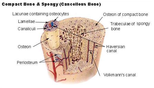
Some, mostly older, compact bone is remodelled to form these haversian systems (or osteons). (on textbook page diagrams note only highlighted labels) compact bone. Concentric circles of bone matrix that make up osteon. The diagram above shows a longitudinal view of an osteon. They are aligned parallel to the long axis of the bone. Good, here in this part, i am going to describe the structure of compact bone. C articular cartilage (a1) c lacuna (b) c collagenous fibers (d) c periosteum (f 1) c compact bone (g) c lamellae (g1) c central canal (h) c osteocyte (i) c canaliculi (j) c perforating canal (k) c blood vessel (l) Concentric lamellae interstitial lamellae central canal lacuna osteocyte canaliculus. This type of bone is located between layers of compact bone and is thin and porous. Compact bone accounts for 80% of the bones in the human body. Osteon model lacunae canaliculi osteocyte. 100x on this image you can see several of the structural units of bone tissue (osteons or haversian systems). The compact bone is the main structure in the body for support, protection, and movement.
There are pores and spaces even in compact bone. Spongy bone is used for more active functions of the bones, including blood cell production and ion exchange. (b) in this micrograph of the osteon, you can clearly see the concentric lamellae and central canals. The light spot is a canal that carries a blood vessel and a nerve fiber. Good, here in this part, i am going to describe the structure of compact bone.
Structure Of Bones Biology For Majors Ii from s3-us-west-2.amazonaws.com These studies show that the internal structure of compact bone changes with age and mirrors its. The canaliculi of mandibular compact bone thinned and developed extensive branching with adulthood but decreased in size and number with advanced age. Trabecular bone, also known as cancellous bone or spongy bone, mainly serves a metabolic function. Compact bone accounts for 80% of the bones in the human body. 100x on this image you can see several of the structural units of bone tissue (osteons or haversian systems). C articular cartilage (a1) c lacuna (b) c collagenous fibers (d) c periosteum (f 1) c compact bone (g) c lamellae (g1) c central canal (h) c osteocyte (i) c canaliculi (j) c perforating canal (k) c blood vessel (l) Diagramme schnell und einfach erstellen. Good, here in this part, i am going to describe the structure of compact bone.
Good, here in this part, i am going to describe the structure of compact bone.
Trabecular bone, also known as cancellous bone or spongy bone, mainly serves a metabolic function. 100x on this image you can see several of the structural units of bone tissue (osteons or haversian systems). Compact bone accounts for 80% of the bones in the human body. Diagramme schnell und einfach erstellen. Concentric circles of bone matrix that make up osteon. Compact bone, or cortical bone, mainly serves a mechanical function. Osteon model lacunae canaliculi osteocyte. Lacunae are minute spaces that contain bone cells, otherwise known as the osteocytes. Spongy bone is used for more active functions of the bones, including blood cell production and ion exchange. Some, mostly older, compact bone is remodelled to form these haversian systems (or osteons). There are two types of bone tissue: Compact and spongy.the names imply that the two types differ in density, or how tightly the tissue is packed together. Good, here in this part, i am going to describe the structure of compact bone.
Compact bone histology slide structure with diagram. It is thick and dense. The compact bone is the main structure in the body for support, protection, and movement. Due to the strong nature of compact bone, compared to spongy bone, it is the preferred tissue for strength. Some, mostly older, compact bone is remodelled to form these haversian systems (or osteons).
Seer Training Structure Of Bone Tissue from training.seer.cancer.gov The canaliculi of mandibular compact bone thinned and developed extensive branching with adulthood but decreased in size and number with advanced age. Formed when an osteoblast becomes embedded in the matrix it has secreted. The series of diagrams below represent the microscopic structure of compact bone tissue. The osteocytes sit in their lacunae in concentric rings around a central haversian canal (which runs longitudinally). The compact bone is the main structure in the body for support, protection, and movement. There are two types of bone tissue: Compact bone accounts for 80% of the bones in the human body. (on textbook page diagrams note only highlighted labels) compact bone.
(b) in this micrograph of the osteon, you can clearly see the concentric lamellae and central canals.
Some, mostly older, compact bone is remodelled to form these haversian systems (or osteons). Trabecular bone, also known as cancellous bone or spongy bone, mainly serves a metabolic function. This is the area of bone to which ligaments and tendons attach. The light spot is a canal that carries a blood vessel and a nerve fiber. The series of diagrams below represent the microscopic structure of compact bone tissue. They are aligned parallel to the long axis of the bone. Compact bone tissue consists of units called osteons or haversian systems. It is thick and dense. Do you want to learn the details of the histology of compact bone with labelled diagram and authentic slide images? Lacunae proceed from the large circular structures of youth to the flat forms of the aged. Good, here in this part, i am going to describe the structure of compact bone. The compact bone is the main structure in the body for support, protection, and movement. This type of bone is located between layers of compact bone and is thin and porous.


0 Komentar